Alnus Rubra and
the Nitrogen-Fixing
Frankia
RESULTS
|
Bob Musgrove
Biology 585
Scanning Electron Microscope
Professor: Dr. Darlene Southworth
Southern Oregon University |
The micrographs on this page are the results of following
specimen preparation method D
which is outlined
in the previous section. The images were scanned with an Hitachi
S-2100 SEM.

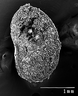
Figure
4. Cross section of a single Alnus rubra root nodule.
at low magnification. Note the 1 millimeter line scale. |
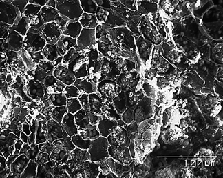
Figure
5. Below is an overview of Alnus rubra root nodule
structure. Note that most of the cell contents (cytoplasm, plastids,
nuclei) appear to have been evacuated during specimen preparation.
Barely visible in this image are individual starch amyloplasts
clustered within the cell walls. The 100 micron line scale represents
0.1 millimeter. |
 |
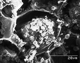
Figure
6. The micrograph on the left illustrates the membrane-bound
vesicles which enclose the Frankia alni. Note how the
interior of the cell appears to be overflowing with vesicles
ranging from 2.5 to 4.5 microns in diameter. |
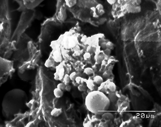
| Figure
7. Frankia vesicles in a Alnus rubra root nodule
specimen different from that used for the above three micrographs.
Note the starch amyloplast, which is 8 microns in diameter,
to the left of the line scale. The line scale represents 0.02
millimeter. |
 |
web page authored by Bob
Musgrove
|


