Alnus Rubra and
|
Biology 585 Scanning Electron Microscope Professor: Dr. Darlene Southworth Southern Oregon University |
MATERIALS Alnus rubra nodules FAA fixative glutaraldehyde osmiun tetroxide phosphate buffer ethanol EMScope Critical Point Drier 750 EMScope Sputter Coater 500 TRI-X 120 film Hitachi-2100 Scanning Electron Microscope Olympus D-620L digital camera standard lab supplies as needed |
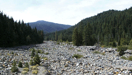
METHODS Alnus rubra root nodules were obtained from a site on the Upper Sacramento River watershed on Forest Service Road 26, 6.3 miles south of the intersection of Hatchery Lane and Old Stage Rd. near Mount Shasta, Siskiyou County , California. The specimens were identified with Peterson's Western Trees (Petrides 1992). Collected specimens were under 3 feet tall and growing in a sand bar in the riparian zone. A garden shovel was used to excavate no more than two feet of root, and vigorous-appearing nodlues were chosen. The whole nodules were stored in river water while they were transported to Southern Oregon University.
A preliminary viewing with the SEM determined that the specimen prepared by Method D, would be the best for imageing the Frankia. Micrographs were taken at various voltages (5kV to 20kV) and magnifications (30x to 2000x). The negatives were then scanned into Adobe Photoshop at 220% of the original size. The web-ready graphics are 72 dpi, 8-bit grayscale GIFs. This website was authored with Adobe Pagemill 3.0.
|
web page authored by Bob Musgrove
